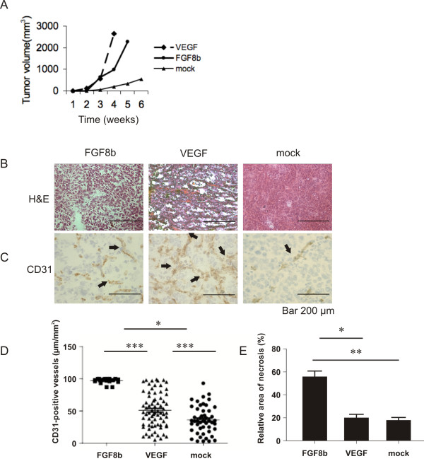
Figure 2.
Growth and morphology of FGF8b, VEGF and mock tumours. A, Growth of subcutaneous FGF8b, VEGF and mock tumours (n = 30, n = 22 and n = 14, respectively). Tumour diameter in 2 perpendicular dimensions was measured once a week and tumour volume was calculated according to the formula V = (π/6)(d1 × d2)3/2 and presented as a function of time (mean ± SEM). Differences in tumour volumes between the groups were significant at all time points between 2 and 4 weeks (p < 0.001). B, H&E staining of representative FGF8b, VEGF and mock tumours (Bar 200 μm). C-D, The density of CD31-positive blood capillaries (μm/mm2) was counted in a blinded manner from 3 fields of the FGF8b, VEGF and mock tumours (51 ± 27 μm/mm2, n = 18, 97 ± 4 μm/mm2, n = 72, and 36 ± 21 μm/mm2, n = 49, respectively), p* < 0.05, p*** < 0.001 (Bar 200 μm). E, The relative area of necrosis was counted in a blinded manner from 3 fields in FGF8b (n = 6), VEGF (n = 6) and mock (n = 6) tumours, p* < 0.05, p** < 0.01.

