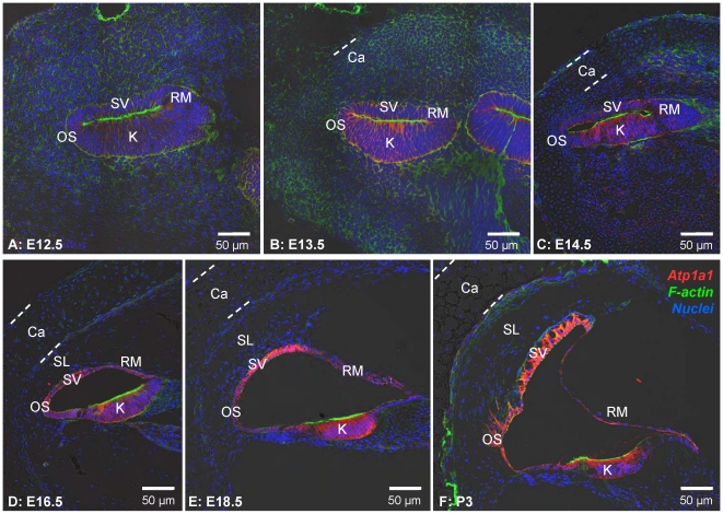
Figure 2. Cochlear development from E12.5 to P3 in Slc26a4+/− mice.
Na+/K+ ATPase (red) was visualized by immunocytochemistry. F-actin (green) and nuclei (blue) were labeled. A-F: Six different stages of development ranging from E12.5 to P3 are shown. Abbreviations: SV, stria vascularis; OS, outer sulcus; K, Kölliker's organ; RM, Reissner's membrane; Ca, otic capsule; SL, spiral ligament. The thickness of the otic capsule is marked by dashed lines.

