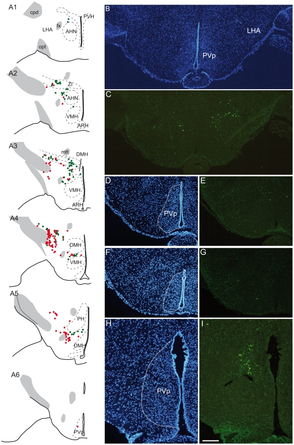
Figure 1. Distribution of MCH perikarya in the mouse hypothalamus.
(A) Distribution of MCH/CART-positive and MCH-positive/CART-negative neurons, (respectively represented by green and red dots) on a series of line drawings of coronal sections through the mouse hypothalamus and arranged from rostral to caudal (A1 to A6). (B–G) Photomicrographs to illustrate the distribution of caudal-most MCH perikarya labeled by the sMCH-AS (C, E, G) at the level of the posterior periventricular nucleus (PVp). Sections were counterstained with DAPI to precisely identify cytoarchitectonic borders of the nucleus (B, D, F). Very few MCH perikarya are seen within this nucleus. (H–I) By contrast in rat, a group of MCH perikarya is labeled within the borders of the PVp. These neurons form a clear cell condensation. AHN: anterior hypothalamic nucleus, ARH: arcuate nucleus hypothalamus, cpd: cerebral peduncle, DMH: dorsomedial nucleus hypothalamus, fx: fornix, LHA: lateral hypothalamic area, mtt: mammillothalamic tract, opt: optic tract, PH: posterior hypothalamic nucleus, PVH: paraventricular nucleus hypothalamus, PVp: posterior periventricular nucleus hypothalamus, VMH: ventromedial nucleus hypothalamus, V3: third ventricle, ZI: zona incerta.

