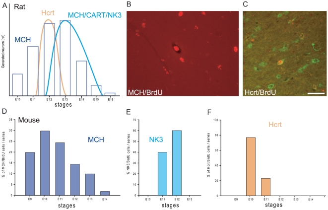
Figure 4. MCH neuron genesis in the mouse hypothalamus.
(A) Histogram summarizing precedent findings in the rat concerning the genesis of MCH, MCH/CART/NK3 and Hcrt neurons in the hypothalamus [12],[18]. (B–F) A similar study combining BrdU, immunohistochemistry and in situ hybridization was conducted in mouse to compare the birthdates of MCH and Hcrt neurons. (B) Photomicrograph illustrating a neuron double-labeled for BrdU (red immunohistochemistry) and MCH (in situ hybridization). (C) Dual immunofluorescence to identify Hcrt (green) and BrdU in the same neurons. Only neurons displaying a densely BrdU-labeled nucleus were taken into consideration. (D–F) Histograms of MCH/BrdU, NK3/BrdU and Hcrt/BrdU neurons. MCH/BrdU positive neurons are seen from E9 to E14, with a peak at E10 (D). NK3/BrdU positive cells in the zona incerta/LHA were observed only at E11 and E12 (E). On the same material, double labeled Hcrt/BrdU perikarya were observed mainly at E10 (F). LHA: lateral hypothalamic area.

