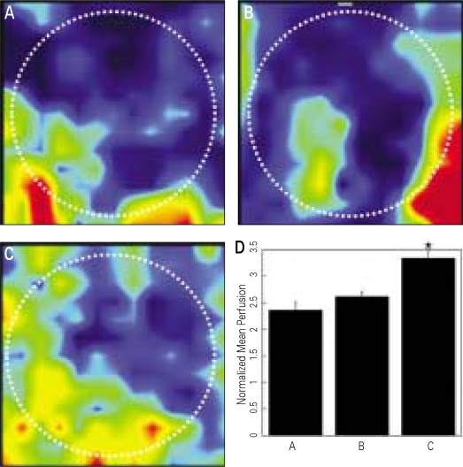
Fig. 3.
A-C: Laser Doppler perfusion images of periosteal region of the cranial defect at 2 weeks after graft procedures. A. β-TCP only, B. β-TCP and conventional PRP activated with 142.8 U/ml thrombin and 4.3 mg/ml CaCl2, C. β-TCP and angiogenic factor-enriched PRP (PRP activated with shear force, 20 mg/ml collagen, 10 U/ml thrombin and 2 mM CaCl2, and containing peripheral blood mononuclear cells), D. Mean perfusion of each group (P < .05). The mean blood flows are normalized to the perfusion measured in the tail of the same animal (unit: voltage). White dotted circles manifest the original defects. Increased blood flow was indicated as color change from dark red to navy blue.

