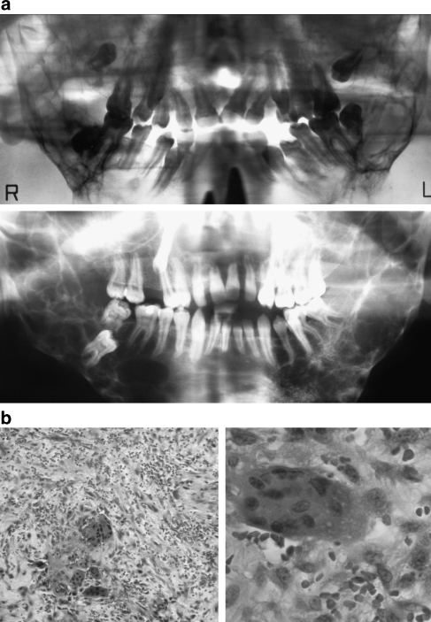
Figure 2.
(a) The radiographs of patients 1 and 4 show bilateral extensive radiolucent lesions in the posterior mandible. (b) The histology pictures of patient 7 show multinucleated giant cells within a fibrous stroma. (HE stain, × 100 and × 400 original magnification. Patients are numbered according to Table 2.)

