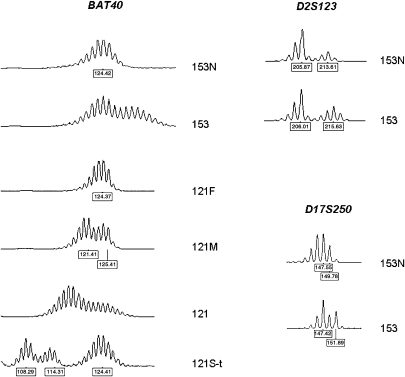
Figure 4.
Type A MSI at the polymorphic mononucleotide repeat marker BAT40 (cases 153 and 121) and at dinucleotide markers D2S123 and D17S250 (case 153). 153N: proband from family 153, normal brain tissue; 153: proband from family 153, glioblastoma; 121F: father, peripheral leukocytes; 121M: mother, peripheral leukocytes; 121: proband from family 121, glioblastoma; 121S-t: sister, colon cancer. Numbers at the bottom of each electropherogram indicate size (in bp).

