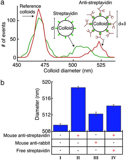
Figure 3.
(a) A histogram showing the distribution of colloid diameters measured from a solution that contains only the reference and streptavidin colloids (green line) and a solution that contains both types of colloids and 0.1 mg/ml monoclonal antistreptavidin antibody (red line). The specific binding of antistreptavidin to the streptavidin colloids produces a clear increase in the diameter of the colloids. (b) A summary of the measurements of the mean diameter of the streptavidin colloids when mixed in different solutions. A single experimental run consists of measuring several hundred colloids of each type in one solution; the plotted bars represent the mean diameter extracted from three to five such runs on the same solution, but using different devices. All solutions contained the streptavidin colloids and the reference colloids in a 0.5× PBS buffer (pH 7.3). The presence of additional components in each solution is indicated by a + in the column beneath the plotted bar. Column I shows the mean diameter measured without any protein added to the solution. A 9-nm increase in colloid diameter is seen in the presence of the specific antibody to streptavidin (0.1 mg/ml mouse antistreptavidin, column II); we attribute this to the volume added to the colloid caused by the specific binding of the antibody. The specificity of the probe is shown by the lack of a similar diameter increase in the presence of isotype-matched irrelevant antibody (0.1 mg/ml mouse anti-rabbit, column III); the small diameter increase in this solution can be attributed to nonspecific adhesion. We also perform an inhibition assay, where the specific binding of the antistreptavidin to the colloid is disrupted by the presence of 0.2 mg/ml free streptavidin (column IV) (the presence of free antigen is shown by the decrease in diameter compared with the antigen-free solution, column II). The error bars in all figures represent the uncertainty in determining the mean diameter based on one standard deviation of the measured distributions. The dominant source of error in our measurements is the intrinsic distribution in the streptavidin colloids' diameter, with smaller contributions from the spread in diameter of the reference colloids and the electrical noise in the current measurement.

