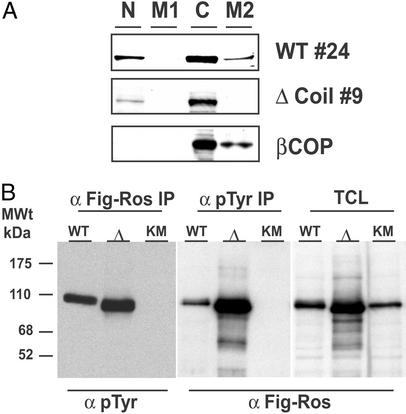
Figure 5.
The Δcoil FIG-ROS mutant displays kinase activity and does not sediment with a Golgi membrane-containing fraction. Cell extracts from retrovirus-infected Rat1 clones were subjected to differential centrifugation cellular fractionation followed by Western blotting with an anti-FIG antibody as described in Materials and Methods (A) and immunoprecipitated with the indicated antibodies and Western blotted for the presence of phosphotyrosine residues (pTyr) and FIG-ROS as indicated (B). N, nuclear and unlysed low-speed fraction; M1, intermediate-speed plasma membrane-containing fraction; C, cytosolic fraction; M2, high-speed Golgi membrane-containing fraction; IP, immunoprecipitate; TCL, total cell lysate; Δ, Δcoil FIG-ROS isoform; KM, K511M FIG-ROS isoform.

