Figure 2.
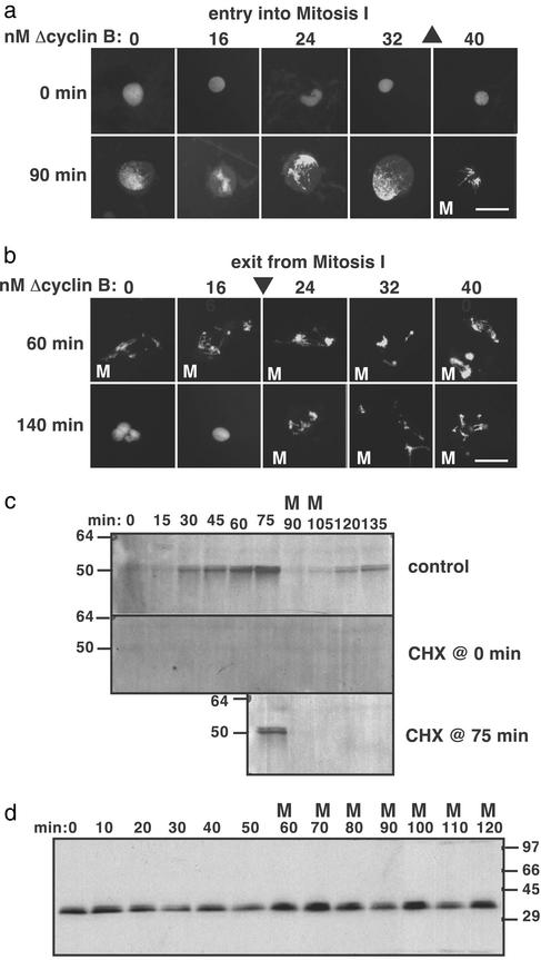
The threshold concentration of cyclin B to enter mitosis is higher than the threshold to exit mitosis. Cycling egg extracts in interphase of cycle 1 were supplemented with Δcyclin B (at t = 0). (a) To measure the activation threshold, CHX was added immediately (t = 0). (b) To measure the inactivation threshold, CHX was added 60 min later when the extract was in mitosis. Fluorescence micrographs of sperm nuclei are depicted. Triangles denote activation threshold (▴) and inactivation threshold (▾) concentrations. (Scale bars = 50 μm.) (c) Extracts prepared as in a and b without exogenous cyclin were immunoblotted for endogenous cyclin B1. (d) A CHX-treated CSF-released extract was supplemented with 150 nM Δcyclin B during interphase (t = 0). Samples were collected and blotted for Δcyclin B. Extracts are labeled M when >90% nuclei on a slide appear mitotic (condensed chromatin, no nuclear envelope). In unlabeled extracts, >90% nuclei were in interphase. Migration of molecular mass standards (in kDa) is indicated.

