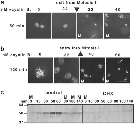
Figure 4.
The threshold concentration of cyclin B to enter mitosis I is higher than the threshold to exit meiosis II. (a) To measure the cyclin threshold for exit from meiosis II, CSF extract was supplemented with CHX and Δcyclin B, then released from CSF arrest by addition of calcium (at t = 0) and photographed under fluorescence microscopy at 50 min. (b) To measure the cyclin threshold for entry into mitosis I, Δcyclin B was added to CHX-treated CSF-released extract at 50 min (when extract was in interphase), and nuclei were photographed at 120 min. Thresholds in a and b were measured in the same extract preparation. Triangles denote threshold concentrations. (Scale bar = 50 μm.) (c) Extracts prepared as in a and b, without exogenous cyclin, were immunoblotted for endogenous cyclin B1.

