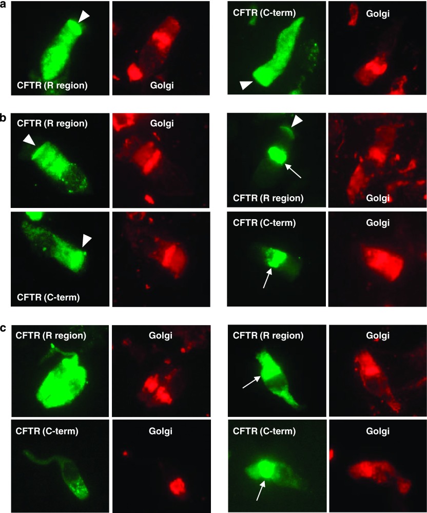
Figure 3.
Immunocytochemical analysis of CFTR in nasal epithelial cells derived from controls (a), from F508del/F508del patients (b), and from F508del/3905insT patients (c). CFTR was detected using either an antibody directed against the C-terminal or the R region of the protein, and the Golgi compartment was stained using wheat germ agglutinin. Arrowheads indicate CFTR localized at the apical membrane of the cells, whereas arrows display an unspecific Golgi-like structure earlier mentioned in the literature.

