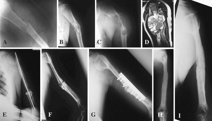
Fig. 2.

Case 2: A 23-year-old male patient was diagnosed with ABC of the humerus. a Anterior posterior (AP) plain radiograph of the left humerus showing pathological fracture of the shaft of the humerus. b AP plain radiograph of the left humerus showing the pieces of the fibular graft at the site of the fracture. c AP plain radiograph of the left humerus showing a large expansile cystic lesion causing total destruction of the bone with ballooning effect. d Coronal T2 MRI views of the left humerus showing multiple fluid levels with hemorrhage inside the cyst. e AP plain radiograph of the left humerus in the immediate postoperative period showing the graft in situ fixed proximally and distally. f AP plain radiograph of the left humerus after 18 months showing healing in the proximal part of the graft and non-union at the distal part. g AP plain radiograph of the left humerus after open reduction and internal fixation (ORIF) with addition of bone graft. h AP plain radiograph of the left humerus after removal of metal showing incorporation of the graft proximally and distally. i AP plain radiograph of the left humerus 8 years postoperatively showing good consolidation, hypertrophy and remodeling of the graft
