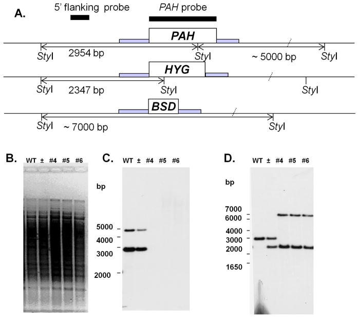
Fig. 3. Generation of L. major PAH null mutants.
(A) Restriction map of the native PAH locus and planned HYG and BSD replacements. The position of StyI sites and predicted fragment sizes are shown. Hybridization probes are shown by black bars, codon regions by open boxes, and regions contained within the targeting fragment by gray bars. (B–D) Southern blot analysis of wild type (WT), heterozygous (±) and PAH null mutant lines (#4, 5, and 6). DNA was subjected to Southern blot analysis using the radiolabeled PAH ORF (panel C) or a 5′ flanking (panel D) probes shown in panel A. Panel B shows the ethidium bromide stained gel prior to transfer.

