
Figure 4.
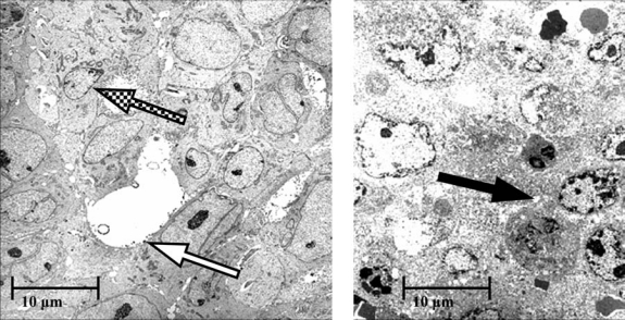
Electron micrographs of human xenografts excised from each of 2 mice, 1 mouse 3 days after intravenous injection of the bioprobes labeled with indium-111 radioisotope (left) and the other similarly treated, but an additional day after the bioprobes were activated by alternating magnetic frequency for 20 minutes (right). The electron micrograph on the left shows healthy breast cancer cells with cell and nuclear membrane outlines (checkerboard arrow) and intracellular cytoplasmic granules. Iron bioprobes located on the cell membrane can also be seen (white arrow). The electron micrograph on the right shows necrotic cell death manifested by the loss of membrane definition and cytoplasmic vacuoles (black arrow).