Figure 2.
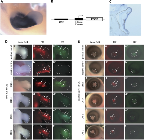
Functional analysis of upstream conserved non-coding elements (CNEs) in chicken limb bud and eye. (A) Shox expression pattern in developing chicken limb (HH26). Whole-mount in situ hybridization shows a strong and specific staining pattern excluding the most distal third of the limb bud. (B) Schematic representation of the vector constructs used to analyze enhancer activity of CNEs in chicken embryos. (C) Shox expression in developing eye (HH26) with specific staining in the cornea. (D) Results of the enhancer assay in chicken limb bud. Pictures show whole mounts of stage HH26 chicken limbs. RFP expression (red) indicates the area electroporated. GFP signal (green) indicates enhancer activity of the tested CNE. A vector containing an SV40 enhancer served as a positive control (a′′, note GFP expression), an empty Bg-EGFP vector served as a negative control (b′′, note lack of GFP expression despite good RFP expression). GFP was detected for three of the four tested CNEs (c′′, e′′, f′′). (E) Result of the enhancer assay in developing chicken eye. Pictures show whole mounts of stage HH26 chicken eyes, circle outlines the cornea. A GFP signal was only detected for the positive control (a′′). As a further control, enhancer activity of non-conserved non-coding elements with comparable CG content was monitored in chicken limb buds (NCE 1: 661 bp, 41% CG; NCE2: 850 bp, 41% NCE3: 480 bp, 43% CG; NCE4: 600 bp, 47% CG). None of these electroporations (23 in total) resulted in a GFP signal, confirming that enhancer activity of CNEs is necessary to evoke a positive signal (control data published in Sabherwal et al10).

