Figure 1.
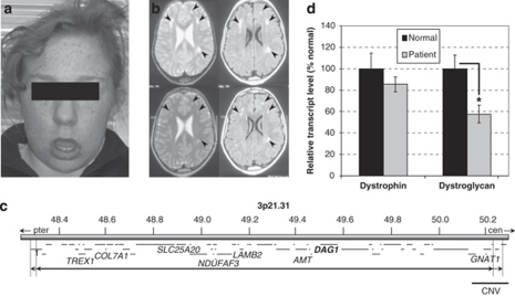
(a) Facial features of the patient, aged 16 years, showing facial hypotonia with everted lower lip and protruding tongue. (b) Axial magnetic resonance images of the patient's brain at age 5 years (upper panels) and 11 years (lower panels). T2-weighted (left hand panels) and FLAIR (fluid-attenuated inversion recovery; right hand panels) images show mild ventricular dilatation and patchy, non-progressive high signal intensity changes in the subcortical white matter of both cerebral hemispheres (arrowheads), relatively symmetrical and slightly more marked in frontal regions. (c) Overview of the deleted region, del(3)(p21.31p21.31)(48286183–50219661)dn (solid line – minimum deletion; dashed lines – maximum deletion). The 65 genes (short horizontal lines) are listed in detail in Supplementary Table 4. The DAG1 gene (bold), seven genes implicated in human genetic disease, and a putatively benign CNV are labelled. (d) Levels of dystrophin and dystroglycan transcripts in normal and patient muscle. Columns show means of three measurements (±SD) normalised to α-dystrobrevin and with the normal muscle expression level set to 100%. *P<0.01.

