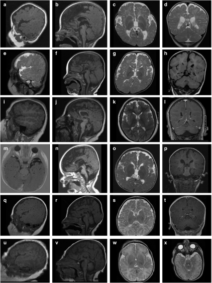
Figure 4.
Brain magnetic resonance images (MRIs) of patients 1–7, excluding patient 4 (fetus). Images a–d of patient 1 at age 6 months, images e–h of patient 2 at age 9 months, images i–l of patient 3 at age 7 years 3 months, images m–p of patient 5 at age 15 months, images q–t of patient 6 at age 3 months, images u–x of patient 7 (sister of patient 6) at postnatal day 1. A suitable parasaggital image was not available for patient 5. In images a, e, i, q, and u, the polymicrogyria around the frontal lobes, and in images a, e, i, and q, the wider-than-normal sylvian fissure is apparent. The sagittal sections b, f, j, n, r, and v show a consistently thin corpus callosum, shortened at the rostrum and genu (with the exception of patient 3, image j), cerebellar hypoplasia in patient 3 (j), and mild cerebellar vermis hypoplasia in patients 5– (images n, r, and v, respectively). The brainstem appears normal in all the patients. Increased subdural spaces are particularly apparent at the temporal poles, as shown in patients 5 and 7 (images m and x, respectively).

