Figure 2.
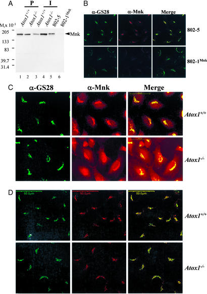
Immunolocalization of Menkes ATPase in Atox1-deficient cells. (A) Immunoblot analysis of Menkes protein in mouse fibroblasts. Cell lysate (100 μg) was separated by 4–20% SDS/PAGE and probed with Menkes antisera (1:2,000) generated against the C terminus of the Menkes ATPase, followed by chemiluminescent detection. Less than 2% of Menkes protein was detected in 802-1 cells compared with wild-type 802-5 cells. (B) Double label indirect immunofluorescence localization (×63) in 802-5 and 802-1 cells grown in basal media, fixed, and stained with GS28 (α-GS28, Alexa 488) and Menkes (α-Mnk, Alexa 546) antibodies, and analyzed by using epifluorescence microscopy. Images of Menkes and GS28 Golgi marker are merged to depict overlapping regions. (Scale bar, 50 μm.) (C and D) Immortalized Atox1+/+ and Atox1−/− cell lines were grown in basal media (C) or in the presence of 200 μM BCS (D) and processed for indirect immunofluorescence studies as described above by using epifluorescence microscopy. (Scale bar, 50 μm.)

