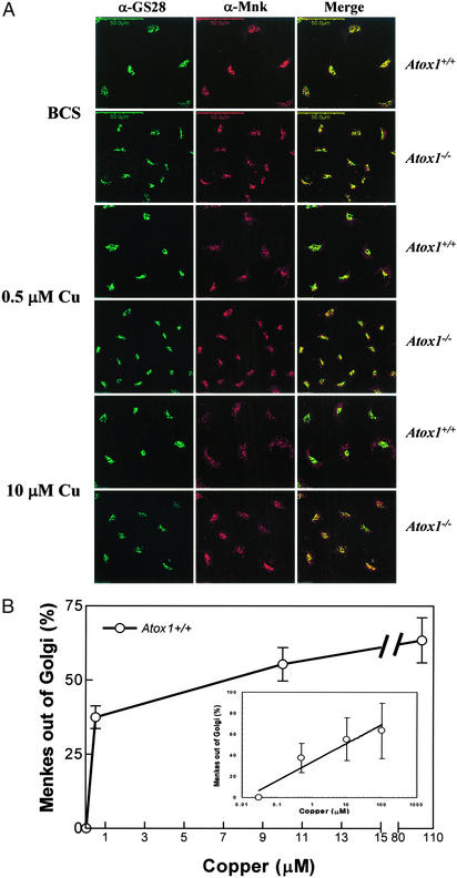
Figure 3.
Characterization of copper-mediated trafficking of Menkes protein. (A) Immortalized Atox1+/+ and Atox1−/− cells were grown on coverslips in the presence of 200 μM BCS for 48 h, followed by the addition of 0.5 or 10 μM CuCl2 for 1 h to the culture media, and processed for double-label immunofluorescence by using Menkes and GS28 antibodies for analysis with confocal microscopy. Images of Menkes and GS28 are merged to depict overlapping regions. (Scale bar, 50 μm.) (B) Quantitative analysis of Menkes signal in the Golgi was measured in immortalized Atox1+/+ cells by using double-label immunofluorescence studies as described in Materials and Methods. Cells were grown in the presence of 200 μM BCS followed by copper treatment for 3 h, and processed for immunostaining. Overlapping signal intensities were analyzed and quantitated by using a laser scanning confocal Olympus microscope and FLUOVIEW FV500 Version 3.3 program. Each data point represents the mean ± SEM from four separate experiments with a minimum of seven scans per experiment per data point. Each scan is comprised of 15–18 cells or at least 105 cells. (Inset) The same analysis plotted onto a logarithmic x axis (μM) for clarity.

