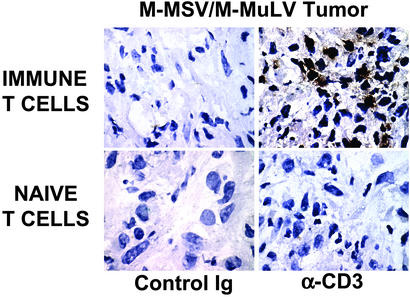
Figure 6.
α-CD3 staining of tumor sections detects greater numbers of immune T cells in the antigen-positive tumor. Shown is staining of tumor sections from animals that bore the M-MSV/M-MuLV tumor. More T cells are detected in the tumor of the animal that received immune T cells than in the animal that received naive T cells. (×1,000.)

