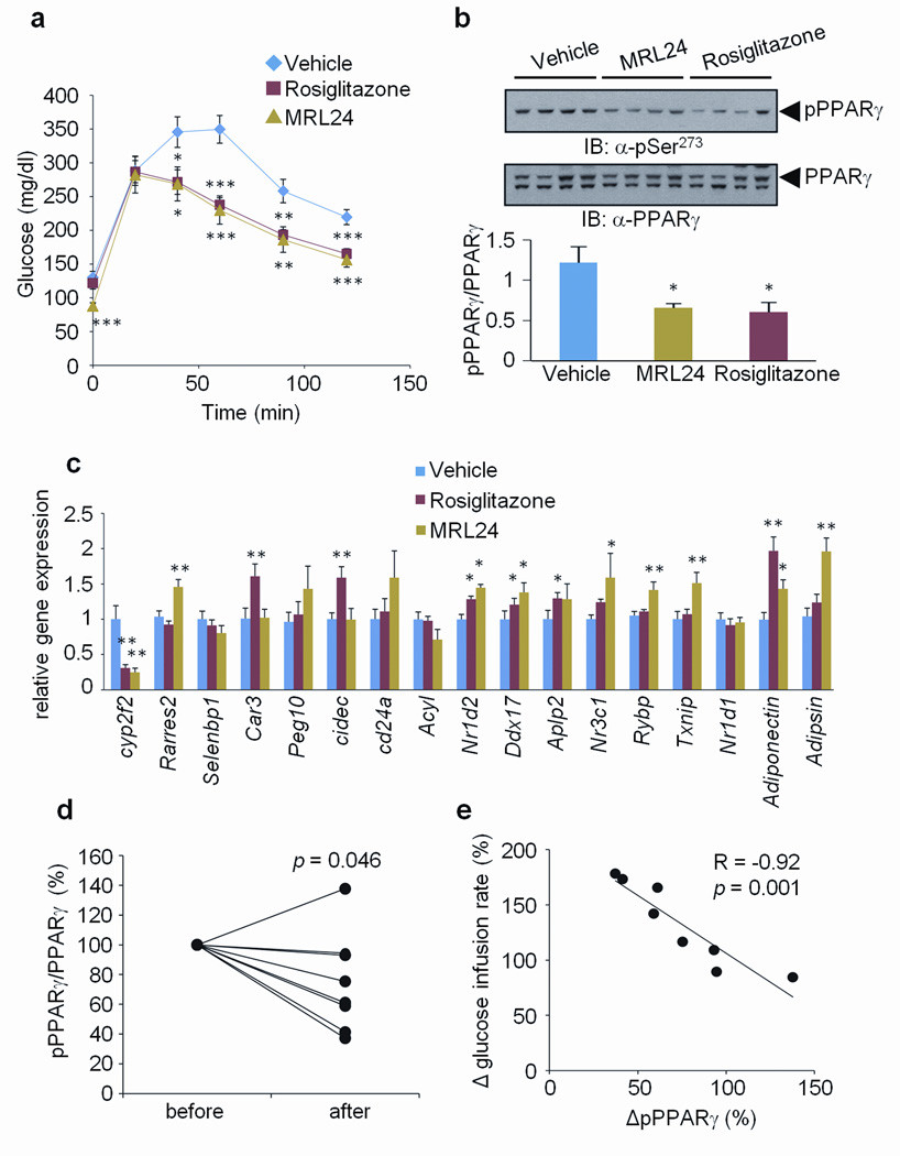
Figure 5. Correlation between the inhibition of phosphorylation and improvement of insulin sensitivity by anti-diabetic PPARγ ligands.
a, Glucose-tolerance tests in 16-week HFD mice treated with vehicle, rosiglitazone or MRL24 (n=10). b, phosphorylation of PPARγ in WAT. c, The expression of gene sets regulated by PPARγ phosphorylation in WAT (Error bars are s.e.m.; *p<0.05, **p<0.01, ***p<0.001). d, Changes of phosphorylation of PPARγ in human fat biopsies which were treated with rosiglitazone for 6 months. before, non-treated; after, 6 months treatment. e, Correlation between the changes of PPARγ phosphorylation normalized to total PPARγ protein and the changes of glucose infusion rate measured by clamp. The data is presented as percent change after 6 months of rosiglitazone treatment.

