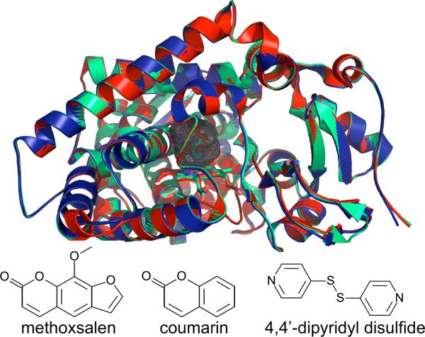
Figure. 2.
A Cα overlay of the methoxsalen (red, PDB ID 1Z11), coumarin (light green, PDB ID 1Z10), and 4, 4'-dipyridyl disulfide (dark blue, PDB ID 2FDY) complexes of CYP2A6. The enzyme requires very little structural rearrangement to bind these ligands. Gray mesh shows the active site cavity of the CYP2A6–coumarin complex and is representative of the cavity of other CYP2A6 structures. Stick diagrams (bottom) show the chemical structures of the ligands bound to each enzyme complex.

