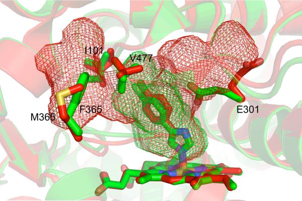
Figure 4.
A comparison of the active site cavities of the 4-CPI complexes of CYP2B6 (red, PDB ID 3IBD) and 2B4 (green, PDB ID 1SUO). Despite their structural similarities (RMSD 0.65 Å), the two enzymes differ with respect to their active site cavity volumes (red and green mesh). The larger cavity in CYP2B6 is created by the movement of E301 out of the active site, the rotations of I101 and V477, and the the change from phenylalanine to methionine at position 365.

