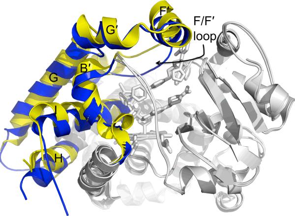
Figure 6.
Cα overlay of ligand free CYP3A4 (blue, PDB ID 1TQN) and the CYP3A4–ketoconazole complex (yellow, PDB ID 2V0M). This ligand free structure is similar to a ligand free structure (PDB ID 1W0E) from another study and the CYP3A4 complexes of progesterone (PDB ID 1W0F) and metyrapone (PDB ID 1W0G). In this overlay, static portions of the enzyme (gray) do not change, but the B', F, F', G', and H helices and the N-terminus of the I helix alter their structures in response to the binding of two ketoconazole molecules. In particular, the F/F' loop changes position by ~5.5 Å.

