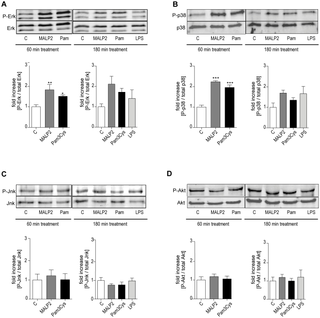
Figure 2. Mitogen activated protein kinase and Akt kinase activation.
After 60 min of perfusion under baseline conditions, isolated mouse lungs were perfused for another 60 or 180 min with Pam3Cys (160 ng/mL, n = 5), MALP-2 (25 ng/mL, n = 5), LPS (1 µg/mL, n = 3) or under control conditions (n = 5). The basal and phosphorylated forms of several kinases were analyzed by immunoblotting after 60 min or 180 min: (A) Erk1/2, (B) p38, (C) Jnk, and (D) Akt kinase. Data were calculated as the ratio of the phosphorylated protein to the total amount of the protein and then referenced to the control on the same gel. Data are shown as mean ± SEM. The micrographs show one representative immunoblot from 3 (LPS) or 5 (C; Pam3Cys, MALP-2) independent experiments.

