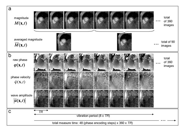
Figure 1.
Diagram for evaluating the complex MRE signal and deducing morphological changes as well as variations in myocardial elasticity during the cardiac cycle. a: Ninety images (t) were obtained from 360 magnitude images M(t) by temporal averaging and used for segmenting the left ventricular cross-sectional area (αLV). b: Three major steps for calculating wave amplitudes U(t) from 360 raw phase images φ(x,t) as described in the text. i) unwrapping, ii) integration, iii) Hilbert transform and display as magnitude U(t).
c: Timing of the image acquisition relative to a vibration period (see text for further details).

