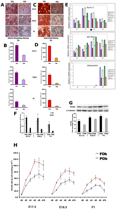
Figure 1. Osteogenic and mitogenic potential of FOb and POb cells.
A, alkaline phosphatase staining performed at day 7 of osteogenic differentiation on frontal osteoblasts (FOb) and parietal osteoblasts (POb) harvested from embryonic 17.5 and 18.5 day (E17.5, E18.5) and post-natal day (P1) mice. B, Alkaline phosphatase enzymatic activity. C, mineralization of extracellular matrix was assessed by Alizarin Red staining at day 21 day. D, quantification of Alizarin red staining. Both alkaline phosphatase activity and mineralization of extra cellular matrix were more robust in FOb than in POb, thus indicating the higher osteogenic capacity of the neural crest derived osteoblasts. E, quantitative real time PCR of osteogenic markers Runx-2, Alk-Phos and Osteocalcin F, cell growth of E17.5, E18.5, and P1 FOb and POb cells was assessed by BrdU incorporation assay at 12 or 24 hours in serum free medium cultures. G, cell growth investigated by immunoblotting analysis of proliferating cellular nuclear antigen (PCNA). Cell lysates were collected after 24 hours culture in serum-free (SF) medium. α-tubulin was used as loading control. Histogram (bottom panel) represents the densitometric analysis of the electrophoresis bands. H, long term growth curve assay performed by cell counting. The values are presented as means ± SD of three independent experiments. BrdU incorporation, PCNA and growth curve assays showed statistical significant higher cell proliferation capability in FOb as compared to POb. The results are presented as the mean ± SD of three independent experiments. Asterisk * represent p<0.05.

