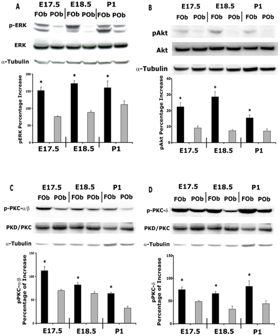
Figure 5. Differential activation of FGF signaling pathways between FOb and POb cells.
A, endogenous phosphorylated ERK protein of MAPK pathway was assessed by immmunoblotting using specific pERK antibody. Total ERK protein levels were analyzed using a pan-ERK antibody. Significantly higher levels of pERK protein were detected in FOb cells compared to POb cells. B, phosphorylated Akt of PI3K pathway was determined with pAkt antibody. Total Akt protein was analyzed with pan-Akt antibody. C, phosphorylated PKC α/β and D, PKC δ of PKC pathway were investigated by immunoblotting analyses using specific antibody against the phosphorylated proteins Total levels of PKC α/β and PKC δ proteins were detected using antibody against non-phosphorylated proteins. FOb cells displayed significantly higher endogenous activation of all three FGF signaling pathways as compare to POb cells. Each membrane was stripped and subsequantially incubated with non-phosphorylated ERK, Akt, PKC α/β, PKC δ and α-tubulin antibody to assess for the total amount of endogenous proteinsand to control for equal loading and transfer of the samples. Histograms represent the densitometric analysis of electrophoresis bands, the relative intensities of bands were normalized to their respective loading control and set as 100% The results are presented as the mean ± SD of three independent experiments. Asterisk * represents statistical significance (*p<0.05).

