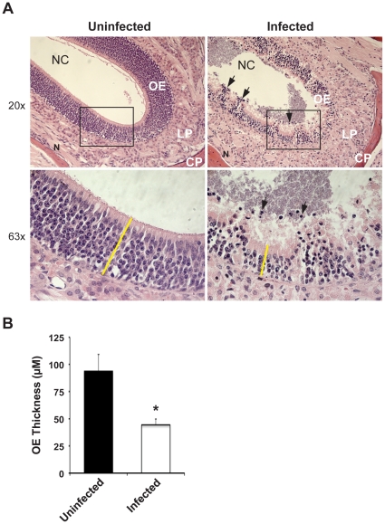
Figure 4. Meningococcal colonization triggers tissue damage of the olfactory epithelium.
(A) 107 CFU of N. meningitidis or PBS (uninfected control) was administrated i.n. to CD46 transgenic mice. At day 3 post-challenge, nasal tissue sections were collected and stained with Hematoxylin and Eosin. Hematoxylin stains negatively charged nuclei dark blue and eosin stains other tissue structures pink. The olfactory epithelium is thereby visualized as a region with a high density of heavily hematoxylin-stained cells. Images show the olfactory epithelial (OE) region of nasal mucosa and the luminal space of the nasal cavity (NC). Epithelial damage and atrophy was induced upon bacterial infection (right panels). Higher magnification (63×) of the box in upper panels is shown in lower panels. Arrows indicate infiltrated cells. Yellow bar: thickness of the OE. Abbreviations: LP, lamina propria; CP, cribriform plate; N, olfactory nerve. (B) Thickness of the olfactory epithelium was measured using a Carl Zeiss Axio Vision 2.05 image analysis system (Zeiss). Upon meningococcal infection, the thickness of the OE is significantly decreased compared to the uninfected mice (A, left panels). *, p<0.05, nonparametric Mann-Whitney test.

