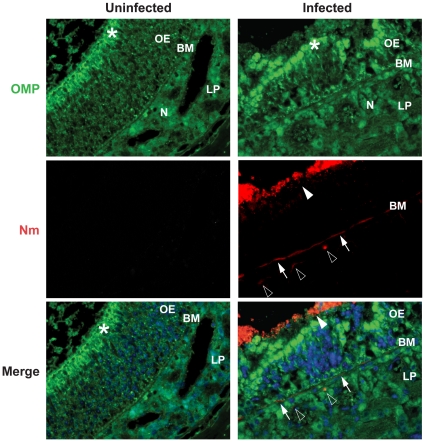
Figure 5. Colonization and invasion of N. meningitidis to the olfactory mucosa.
107 CFU of N. meningitidis were given i.n. to CD46 transgenic mice (Infected). At day 3 post-challenge, nasal tissue sections were collected and stained for N. meningitidis as described in Materials and Methods. Olfactory epithelium (OE) was determined by immunofluorescence staining using an antibody against olfactory marker protein (OMP), which recognizes mature cells of the olfactory sensory neuron and their dendrites in the olfactory epithelium. Strong continuous OMP immunoreactivity (green) could be seen in the apical olfactory epithelium (OE, *) and bundles of olfactory nerves (N) in lamina propria (LP). Nuclei were stained with DAPI. Bacterial signals (Nm, red) were detected on the surface of the olfactory epithelium (arrwohead), at the basement layer (BM, arrows) and in lamina propria (LP, empty triangles). No bacterial staining was found in the corresponding regions of uninfected mice.

