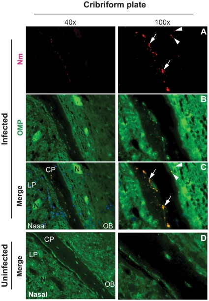
Figure 6. N. meningitidis invades the CNS through the olfactory nerve.
CD46 transgenic mice were challenged i.n. with 107 CFU of N. meningitidis (Infected). At three days post-infection, the head tissue samples were prepared by sectioning through the midsagittal plane to give a complete anatomic structure between the nasopharyngeal region (nasal) and the olfactory bulb (OB). (A) Bacteria (Nm, red signal) were detected in the cribriform plate (arrows) and at the edge of olfactory bulb (arrowheads). (B) Olfactory nerve system was determined by using an antibody against olfactory marker protein (OMP, green signal). (C) In merged images, all bacterial signals co-localized with OMP, a marker for the olfactory nerve. (D) No bacterial staining was found in the corresponding regions of uninfected mouse. Abbreviations: LP, lamina propria; CP, cribriform plate; OB, olfactory bulb; N, olfactory nerve.

