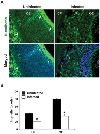
Figure 7. Depletion of N-cadherin in olfactory epithelium upon N. meningitidis infection.
CD46 transgenic mice were challenged i.n. with 107 CFU of N. meningitidis. Mice challenged with PBS were set as uninfected control. At day 3 post-challenge, head tissue was collected and tissue sections were stained using N-cadherin and N. meningitidis antibodies as described in Materials and Methods. (A) In uninfected mice (left panels), strong and continuous N-cadherin staining (green) was identified in the apical layer (arrows) of the olfactory epithelium (OE) and bundles of olfactory nerve (arrowheads) in lamina propria (LP). In infected mice (right panels), the N-cadherin signal was significantly decreased in the corresponding regions. In merged image, bacteria (red) were detected at the olfactory mucosa surface of infected mice. Nuclei were stained with DAPI. (B) Quantification of N-cadherin staining intensity by software Image J. After infection, expression of N-cadherin was significantly decreased in both OE and the LP region. The data are presented as mean ± SD. Significant reductions of signal intensity compared with uninfected mice are indicated with asterisks (*, p<0.05, nonparametric Mann-Whitney test). Abbreviations: OE, olfactory epithelium; LP, lamina propria; CP, cribriform plate.

