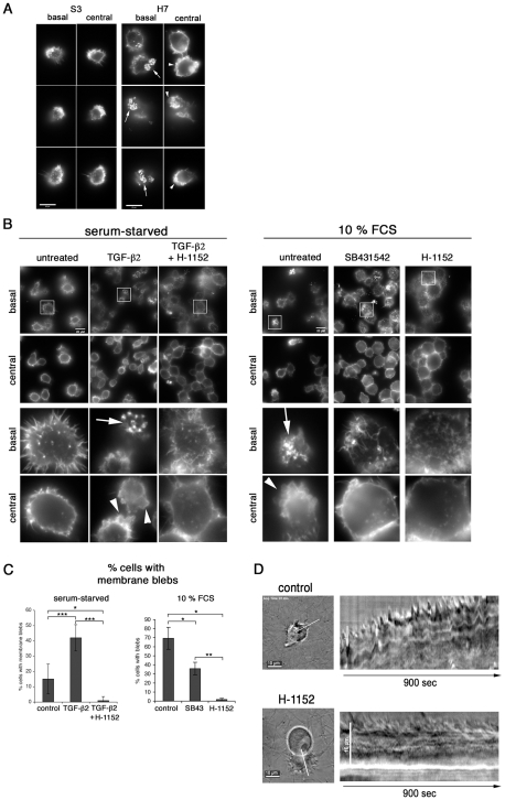
Figure 5. TGF-b promotes podosomal adhesion structures and ROCK-mediated cortical actin-rich membrane blebs.
A, S3 and H7 cells were seeded onto a gelatin/fibronectin matrix. After 24h in culture, the actin cytoskeleton was visualized by IF microscopy. Basal and central focal planes are shown and actin structures were visualized by phalloidin staining at basal adhesion sites and in the lateral cortex, respectively. B, Left panel: H7 cells were serum starved in 0.5% FCS for 24h and then seeded onto a gelatin/fibronectin matrix in starvation medium and in the absence or presence of TGF-b2 (10ng/ml) and H-1152 (10uM), as indicated. Right panel: H7 cells were grown in complete growth medium and then seeded onto a gelatin/fibronectin matrix in complete growth medium (10% FCS) and in the absence or presence of SB431542 and H-1152, as indicated. Analyses was performed as described in A. Magnifications in lower half of the panels are four-fold of areas highlighted with frames. C, Statistical analysis of experiment shown in panel B. Percentages of cells with membrane blebs within total cell populations was determined. Every cell in at least four independent areas per condition was analysed. 0.5% FCS: control: n = 121 (15.11%−/+9.7), TGF-b: n = 235 (42.00%−/+8.5), TGF-b+H-1152 N = 143 (1%); 10% FCS: control: n = 60 (69.27%−/+12.12), SB431542 = SB43: n = 167 (35.84%−/+6.94), H-1152: n = 81 (1.66%−/+1.47). Arrows indicate podosomal adhesion structures, arrowheads membrane blebs. D, Infected cells were embedded in fibrillar collagen and live-cell video microscopy was performed 24h after embedding. Where indicated, 10uM Rho-kinase inhibitor H-1152 was added 1h before acquisition of movies. Sill images of movies and kymographs along the white line indicated are shown.

