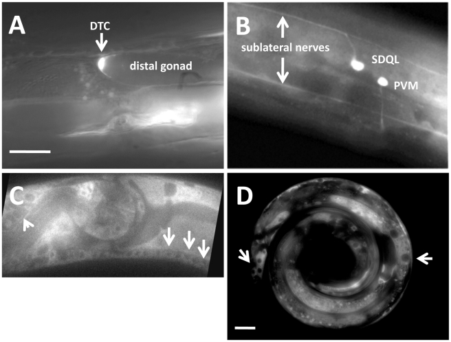
Figure 4. RACK-1 is expressed in most cells, including neurons and the gonadal distal tip cells.
Panels are fluorescent micrographs of animals harboring rack-1::gfp transgenes. (A and B) are fusions of the rack-1 promoter to gfp; and (C and D) are fusions of the entire full-length rack-1 coding region to GFP. A) A distal tip cell expressed rack-1::gfp. B) Neurons expressed rack-1::gfp. C) Full-length RACK-1::GFP was expressed in neurons in the ventral nerve cord (arrows) and in the amphid (arrowhead). D) Full-length RACK-1::GFP was excluded from nuclei in tail hypodermis and gut (arrows). The scale bar in A represents 5 µm for A–C, and the scale bar in D represents 5 µm.

