Figure 1.

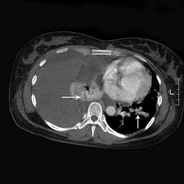
Sample figure title. Contrast-enhanced computed tomographic scan of the thorax shows blood in the right pleural space and compression atelectasis of the right lung. Note also the high-density nodules (arrow) located right lower lobe and left lower lobe, possibly indicative of the PAVMs.
