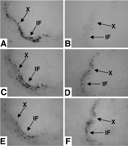
Figure 3.
Colocalization of IRX5, IRX1, and IRX3 as shown by tissue printing. Tissue prints of wild-type (A, C, and E) and irx5-1 (B, D, and F) stems probed with anti-IRX5 antibody (A and B), anti-IRX1 antibody (C and D), or anti-IRX3 antibody (E and F). X, xylem; IF, interfascicular region. Arrows indicate the same points in serial sections.

