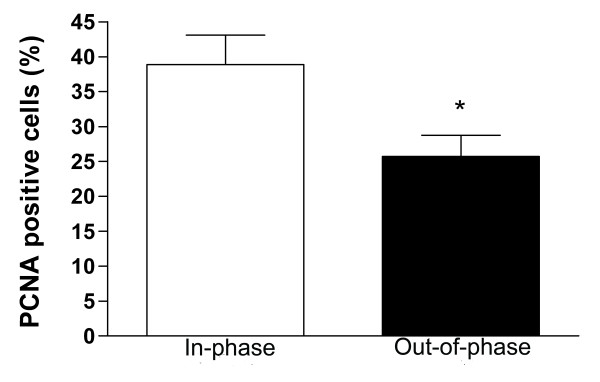
Figure 4.
Cell proliferation in in-phase and out-of-phase endometrium. Histological sections were immunostained for PCNA expression (see text). The total number of epithelial cells in 10 representative fields was counted. Cell proliferation was quantified in the epithelial fraction as percentage of PCNA positive cells. Out-of-phase endometrium shows a significantly lower percentage of PCNA positive cells than in-phase endometrium. * p < 0.05 vs. Control.

