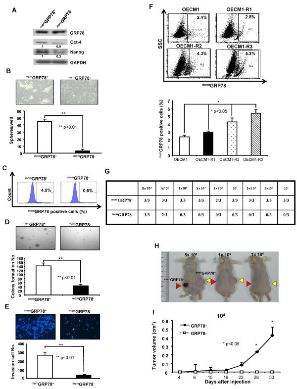
Figure 2.
Cancer stem cells properties of memGRP78+ and memGRP78- HNSCCs in vitro and in vivo. (A) Expressions of pluripotent stemness genes (Oct4 and Nanog) in memGRP78+ and memGRP78- HNSCCs were determined by western analysis. The amount of GAPDH was referred as loading control. (B) Representative images of tumorsphere-forming ability in memGRP78+ and memGRP78- HNSCCs grown under defined serum-free selection medium as described at Materials and Methods. The numbers of spheres were further calculated using microscope. Results are means ± SD from three experiments. (**, p < 0.01) (C) Sorted memGRP78+ and memGRP78- cells were further cultivated with standard medium containing 10% serum. At day 10, the percentage of memGRP78 expression was re-analyzed by flow cytometry. (D) To elucidate the anchorage independent growth, single cells suspension of memGRP78+ and memGRP78- cells plated onto soft agar and analyzed. Results are means ± SD of triplicate samples from three experiments (**, p < 0.01) (E) Invasion ability of memGRP78+ and memGRP78- cells were plated onto transwell coated with matrigel and analyzed. Results are means ± SD of triplicate samples from three experiments (**, p < 0.01) (F) Increased radio-resistance properties (OECM1-R3 > OECM1-R2 > OECM1-R1 > parental OECM1) positively correlates memGRP78 expression in HNSCCs by FACS analysis. (*, p < 0.05) (G) Summary of the in vivo tumor growth ability of different numbers of memGRP78+ and memGRP78- cells examined by xenotransplantation analysis. (H) Representative tumor growth of memGRP78+ and memGRP78- HNSCCs was generated in the subcutaneous space of recipient nude mice (Yellow arrows: memGRP78+ HNSCCs; Red arrows: memGRP78- HNSCCs). (I) Tumor volume was measured after inoculation of memGRP78+ and memGRP78- HNSCCs in nude mice. Error bars correspond to SD (lower panel). (*, p < 0.05).

