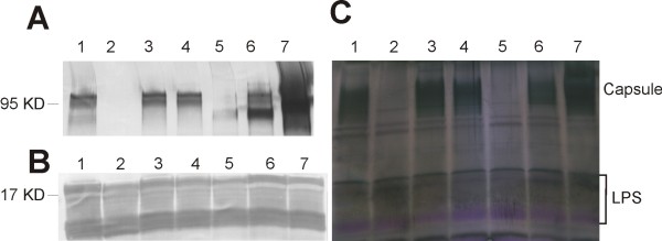
Figure 4.
Immunoblots and stains-all/silver-stain of V. parahaemolyticus. Whole cells lysate treated with DNase, RNase and pronase was separated on polyacrylamide gel, transferred to PVDF membrane and probed with K6 specific antiserum (A), or O3 specific antiserum (B). Total polysaccharides were visualized by stains-all/silver-stain on polyacrylamide gel (C). lane 1, wild type VP53; lane 2, ∆CPS mutant; lane 3, ∆EPS mutant; lane 4, ∆wzabc mutant; lane 5, ∆0220 mutant; lane 6, ∆0220 mutant with trans-complementation; lane 7, ∆VP215-218 mutant.

