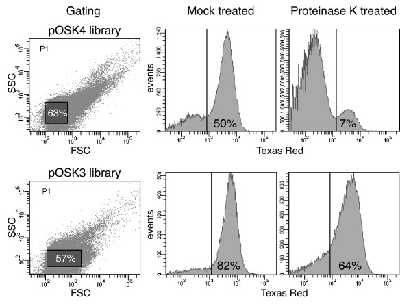
Figure 2.
FACS plots of OspA:mRFP1 mutant populations. Both pOSK4 (pRJS1009-based) and pOSK3 (pRJS1016-based) B. burgdorferi libraries were assayed. The two panels to the left indicate the gating used. Forward scatter (FSC) is plotted against side scatter (SSC). The percentage of events, i.e. cells inside the gated population (shaded rectangles) is indicated. The four panels to the right show the distribution of presorted, i.e. OspA:mRFP1-expressing fluorescent cells upon treatment with proteinase K. Mock treated cells were incubated in buffer only. Fluorescence measured via a Texas Red filter is plotted against number of events, i.e. cells. The vertical line indicates the cut-off fluorescence for sorting. The percentage of events within the fluorescent population is indicated.

