
Figure 3.
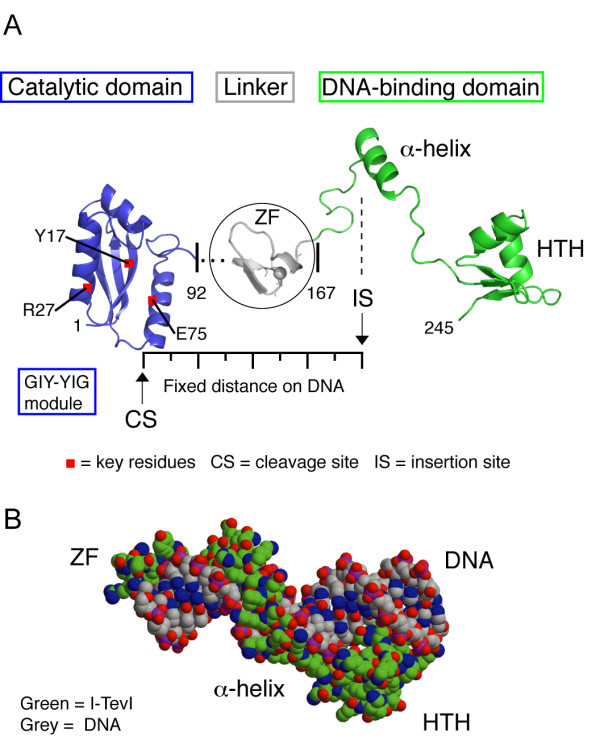
I-TevI structure. A. Two domains of the enzyme joined by a linker. The catalytic GIY-YIG domain (blue) is separated from the DNA binding domain (green) by a 75-amino acid linker, which includes the zinc finger (grey). The DNA binding domain consists of elongated segments, an α-helix and a helix-turn-helix (HTH) module. B. Space filling model of the DNA-binding domain and zinc finger on DNA. The protein is bound to a 20-bp DNA substrate.