Figure 1.
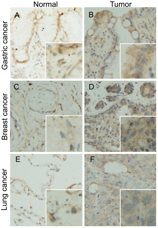
VLDLR II detection by IHC. Representative Immunohistochemical staining of VLDLR II protein in paraffin-embedded human different cancers and matched adjacent noncancerous tissues. A, B: Gastric normal and tumor tissue; C, D: Breast normal and tumor tissue; E, F: Lung normal and tumor tissue. VLDLR II staining show predominantly membranous localization, and a much smaller subset also showed cytoplasmic staining. Normal tissues seen in A, C, E are weak-to-moderate stained, while in cancer cells, VLDLR II staining are strongly intense (B, D, F). Original magnification: × 200; × 400 (insets).

