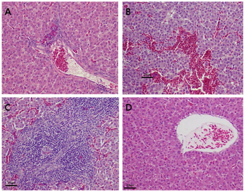
Fig. 3.

Microscopic lesions in the liver. (A) Section of a liver from a chicken inoculated with RNA transcripts from the avian HEV-VA cDNA clone (pT7-aHEV-VA) showing mild periphlebitis. (B and C) Sections of a liver from a chicken inoculated with RNA transcripts from the avian HEV-prototype cDNA clone (pT7-aHEV-5) showing hemorrhages (B) and severe periphlebitis (C). (D) Section of a liver from a negative control chicken inoculated with PBS buffer. The tissues were stained with hematoxylin and eosin.
