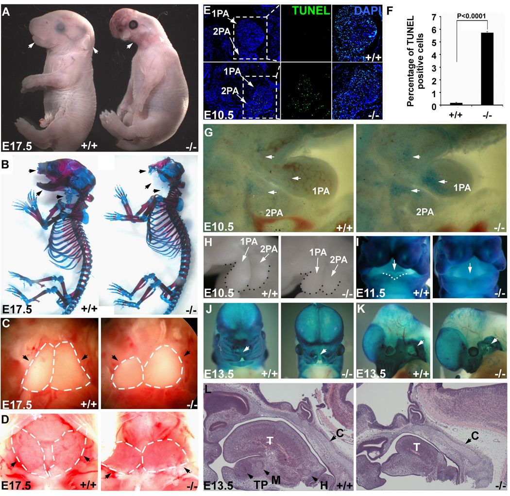
Figure 1. Neural-crest-specific ablation of Dicer resulted in severe craniofacial defects.
In all images, wild type (+/+) control and NCC-Dicer mutant (−/−) embryos are shown. (A) Gross morphology of E17.5 embryos. Neural-crest-specific Dicer mutant embryos displayed severe craniofacial defects (arrows). (B) Skeletal preparation of E17.5 embryos. Most of the neural-crest-derived cartilages and bones, including maxilla, mandible and nasal bones (arrows), were absent in Dicer mutants. (C) Thoracotomies showed the hypoplasia of thymus (arrows) in mutant embryos. (D) Dicer mutant mice exhibited underdeveloped thyroid glands (arrows). (E) TUNEL assay showed an abnormal apoptosis in the 1st (1PA) and 2nd (2PA) pharyngeal arches in mutant embryos. (F) Statistics of TUNEL-positive cells. Three discontinuous sections containing 1st and 2nd pharyngeal arches were stained and counted. (G) Whole mount NBS staining consistently revealed the abnormal apoptosis in mutants. (H) The morphology of 1st and 2nd pharyngeal arches of E10.5 embryos showed the reduced size of pharyngeal arches in Dicer mutants. (I–K) Whole mount β-gal staining of E11.5 and E13.5 embryos containing Rosa-LacZ indicator. E11.5 Dicer mutant embryos failed to form the lateral lingual swellings (I, arrows); E13.5 Dicer mutant embryos displayed a cleft lip (J, arrows) and a hypoplastic pinna (K, arrows). (L) Histological examination of E13.5 embryonic heads. The neural-crest-derived Meckel’s cartilage, hyoid cartilage and tooth primordium failed to form in mutants, while the development of non-neural-crest-derived cartilage primordium of clivus is unaffected. C: cartilage primordium of clivus; H: hyoid cartilage; M: Meckel’s cartilage; T: tongue; TP: tooth primordium.

