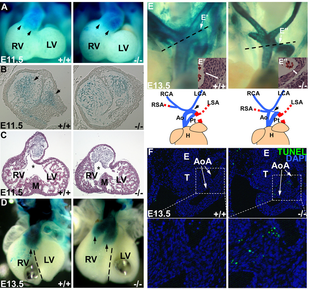
Figure 3. Dicer and miRNAs are required for proper development of neural crest cells.
(A) Whole mount β-gal staining of E11.5 embryos containing Rosa-LacZ indicator showed the migration of cardiac neural crest to the caudal limit of outflow tract (arrows). (B) Histological examination of the stained outflow tract. The Dicer-deficient cardiac NCCs failed to form two columns of condensed mesenchyme in outflow tract. (C) Histological examination of E11.5 heart. The entrance of outflow tract for future aorta (asterisks) had a rightward malalignment with muscular interventricular septum. (D) Whole mount β-gal staining of E13.5 embryonic hearts indicated the rotation defect and malalignment of conus cushion. Two prongs of Dicer-deficient cardiac NCCs (arrows) located in right ventricle in a side-by-side manner. The dash lines indicate the ventricular septum. (E) Neural-crest-derived great vessels in E13.5 embryos were shown by β-gal staining. The blue lines in cartoon indicate the neural-crest-derived vessels, while the red dash lines indicate the vessels not contributed by cardiac NCCs, which cannot be shown in β-gal staining. Mutant embryos displayed a hypoplastic aortic arch artery. Histological examination of aortic arch was performed in the orientation indicated by dashed lines. The regions (E’, E’’) indicated by arrows were shown at bottom right and Supplemental Figure VII. The aortic arch in mutant embryos had a thinner vessel wall with fewer smooth muscle cells. (F) TUNEL assay with E13.5 embryo sections containing aortic arch showed an abnormal apoptosis of neural-crest-derived smooth muscle cells in the vessels in Dicer mutant embryos. (G) Statistics of TUNEL-positive cells. Three discontinuous sections containing aortic arches were stained and counted. Ao: aorta; AoA: aortic arch; E: esophagus; H: heart; LA: left atrium; LCA: left carotid artery; LSA: left subclavian artery; LV: left ventricle; M: muscular interventricular septum; Pt: pulmonary trunk; RA: right atrium; RCA: right carotid artery; RSA: right subclavian artery; RV: right ventricle; T: trachea.

