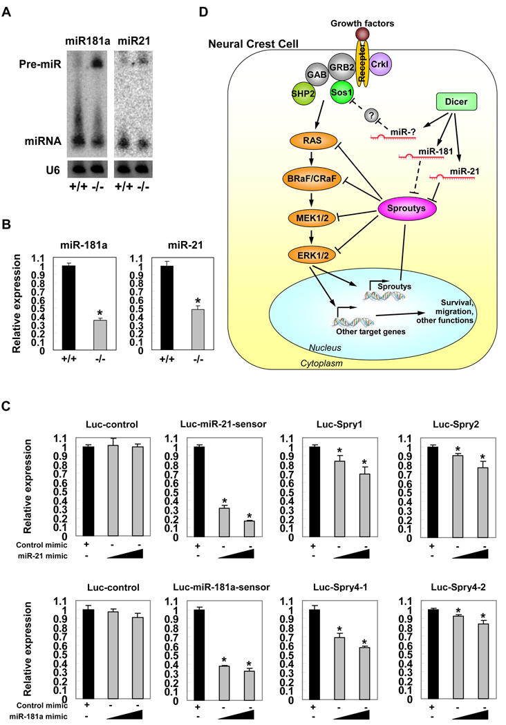
Figure 5. Repression of sprouty expression by miRNAs.
(A) Detection of the haploinsufficiency of mature miRNAs and the accumulation of miRNA precursors in mutant pharyngeal arch tissue samples by Northern blot. (B) Quantitative real-time PCR assays to measure the expression level of mature miR-181a and miR-21 in mutant pharyngeal arch tissues. *: P < 0.01. (C) Hela cells were transfected with indicated luciferase reporters, along with either miR-21 or miR-181a duplex mimic. A Renilla luciferase vector was cotransfected to serve as an internal control for normalization. Cells were harvested and luciferase activity measured 24 hours after transfection. Values are presented as mean luciferase activity ± SD relative to the luciferase activity of reporters with control duplex mimic. *: P < 0.05. (D) Dicer and miRNAs are suggested to participate in a regulatory cascade to modulate the expression levels and activities of MEK/ERK signaling pathway in neural crest cells.

