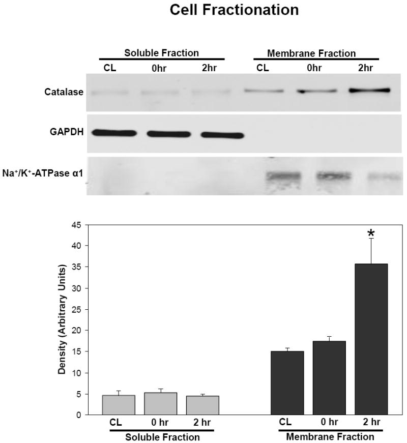
Figure 2.

Subcellular localization of catalase after 2 hr incubation with 0.5 uM catalase-SKL. After incubation, myocytes were fractioned into standard soluble and membrane fractions and probed for catalase using SDS-PAGE and western blot techniques. Upper panel: GAPDH used a marker for the soluble fraction and Na+/K+ATPase used as a marker for the membrane fraction to ensure equal loading. Lower panel represents quantitation of the catalase western blot in the upper panel. At zero time, there were no significant differences between control (CL) and treated myocytes in terms of distribution. After 2 hr of incubation however, virtually all the catalase was localized in the membrane fraction in treated myocytes consistent with a peroxisomal localization of the transduced enzyme. Significantly different from control; *p<0.05. Similar results were seen in three experiments.
