
Figure 4.
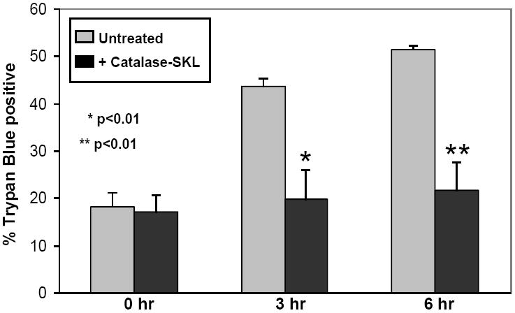
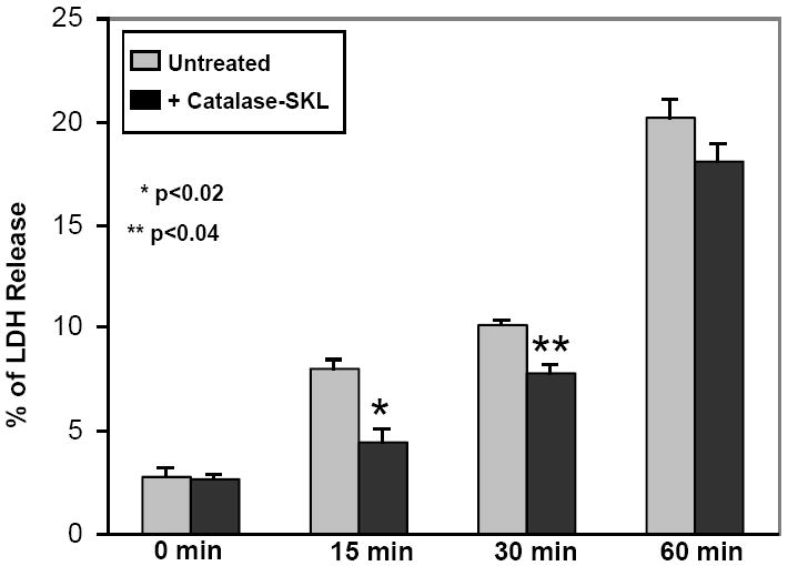
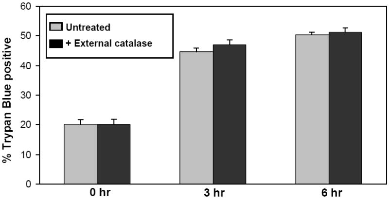
Effect of catalase-SKL on cell death. 4A. Control and catalase-treated (0.5 uM) NRVM were subjected to 3 or 6 hr of hypoxia followed by 1 hr of reoxygenation and cell death was assessed using trypan blue permeability. Compared to control myocytes, treated cells sustained substantially less death indicating a strong cardioprotective effect (* p≤ 0.01 vs. control; ** p< 0.01 vs. control. n=3 for each). 4B: Control and catalase-SKL-treated NRVM were subjected to 15, 30, or 60 min of simulated ischemia followed by 1 hr of reperfusion and cell death was assessed using LDH release into the culture media. At 15 and 30 min of MI/R, treated myocytes sustained substantially less cell death indicating a strong cardioprotective effect (*p≤ 0.01 vs. control; ** p< 0.01 vs. control. n=3 for each). However, after severe ischemia (60 min) the protective effect was largely lost (p=not significant; n=3). 4C: Control myocytes were compared to myoyctes treated with (0.5 uM) non-transducible (i.e. extracellular) catalase (labeled NT catalase). Myocytes were subjected to 3 or 6 hr of hypoxia followed by 1 hr of reoxygenation and cell death was assessed using trypan blue permeability. Extracellular catalase was present throughout the hypoxic incubation but failed to protect against cell death after either 3 or 6 hr of hypoxia/reoxygenation. (p=not significant; n=3 for each group).