
Figure 1. Ultrasound analysis of vascular remodeling in WT and Bmal1-KO mice.
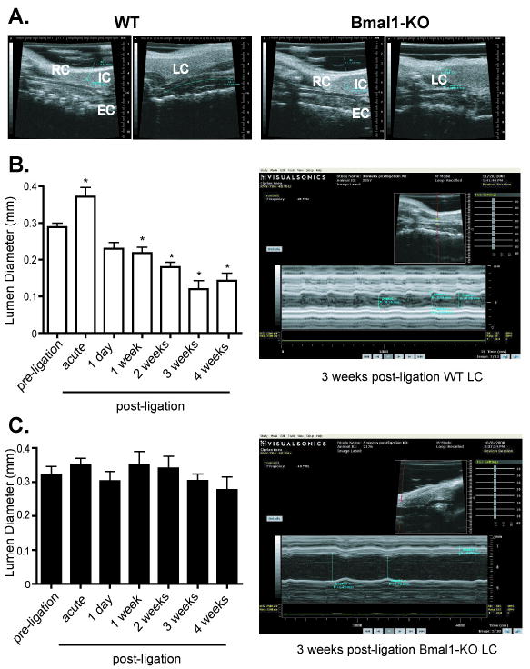
A. The common carotid artery was visualized through echo ultrasound imaging using B Mode (see methods). Typical vascular remodelling evident as a lumen reduction was evident after 4 weeks of arterial ligation in male, wild-type mice (left two panels), Bmal1-KO mice exhibited no change in diameter or a paradoxical increase diameter (right two panels). In the unligated RC, the EC and IC were clearly visible, but absent in the ligated LC in WT and Bmal1-KO mice. B. Ultrasound imaging using M-Mode (see methods) was used to observe the common carotid artery of WT mice after 1 day to 4 weeks of ligation. End diastolic lumen diameter was measured as an index of live in vivo vascular remodelling. Even after 1 day of arterial ligation, a reduction in lumen diameter was observed that progressed through 4 weeks post-ligation in WT mice (right top panel). C. Bmal1-KO mice exhibited no change in diameter (right bottom panel) (N= 6/ group, *p<0.05 by one-way ANOVA versus corresponding preligation lumen diameter)